
Tissue Dissection
Click on the links below to browse HBSFRC’s Protocols:
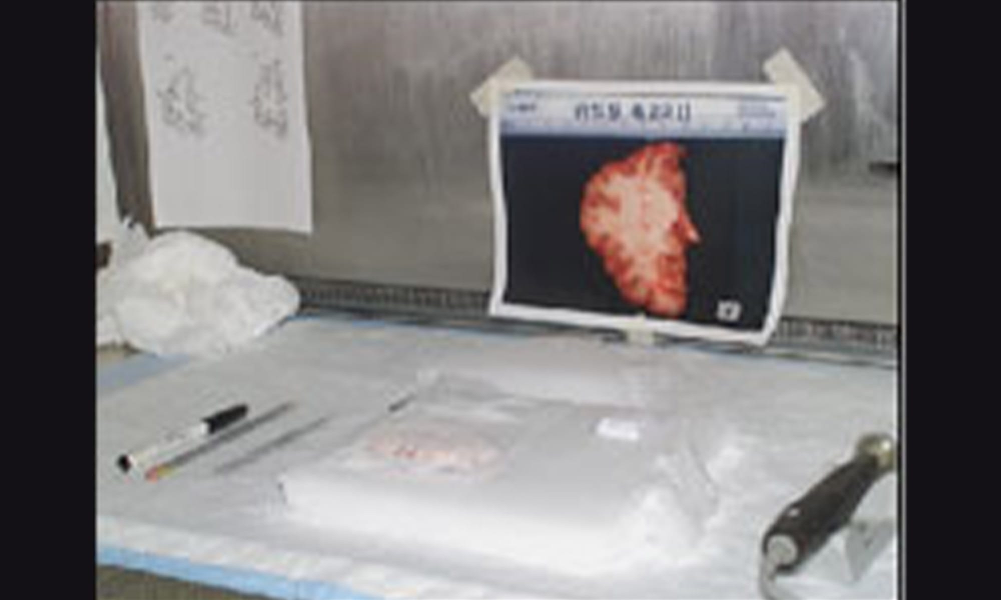
The slab is ready for dissection
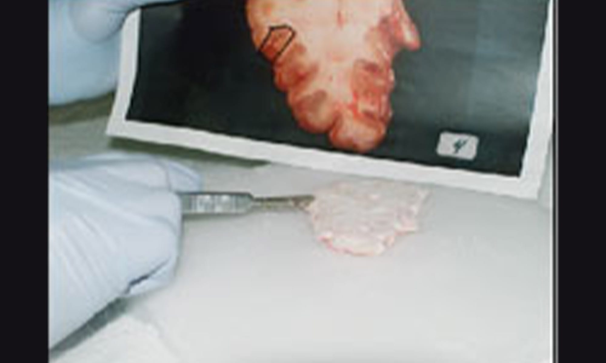
The digital image provides the roadmap for the dissection of the frozen slab
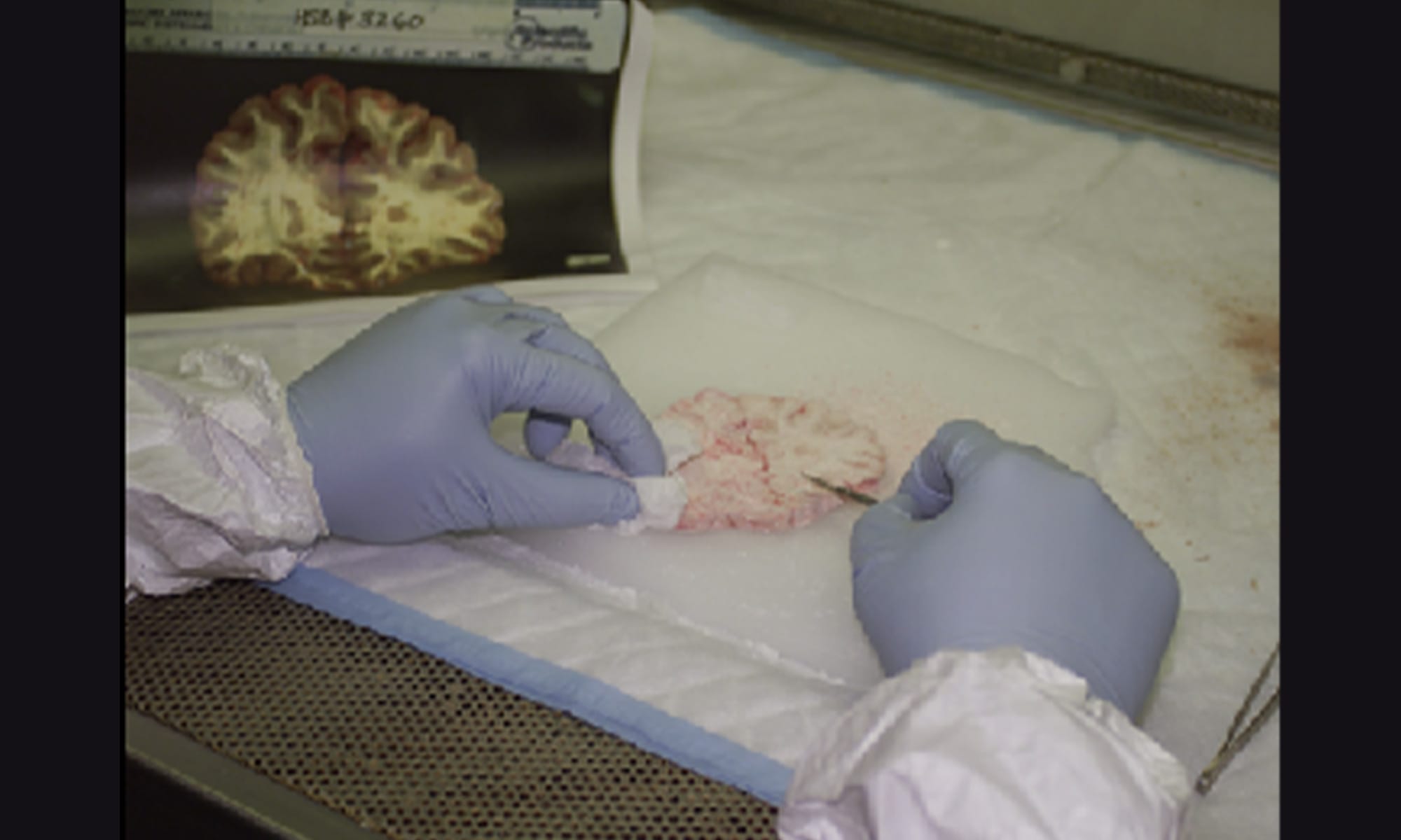
The slab is cleaned
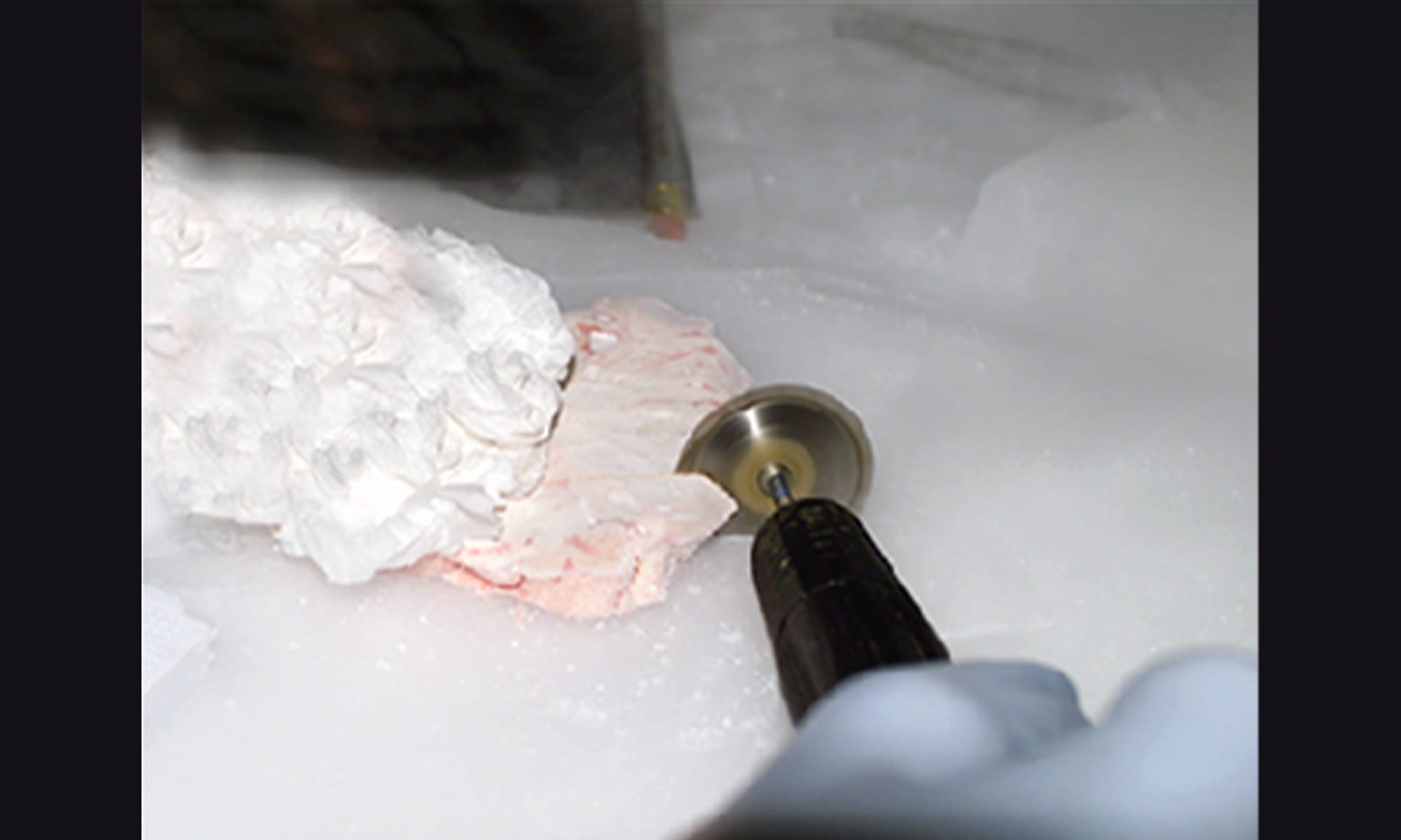
The slab is dissected with a saw. The saw is chilled while dissecting
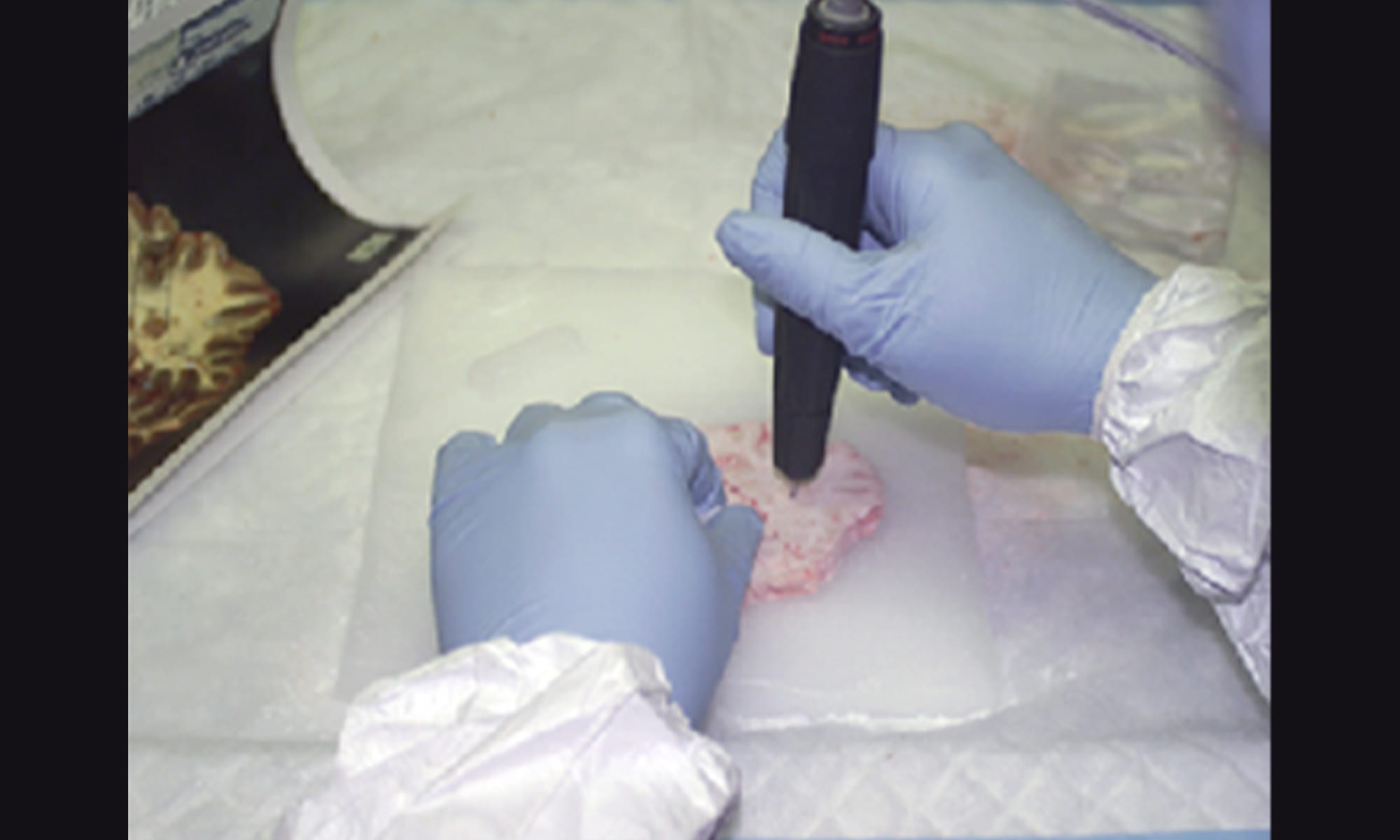
Ultrafine dissection is performed with a dental drill
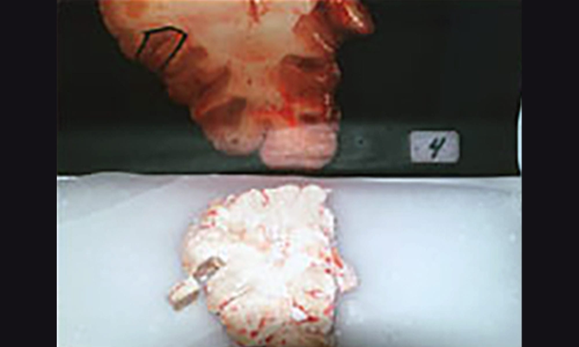
The final drill dissected slab


